


X-ref For other Roundups in this issue that cross-reference with Foot & Ankle see: Research Roundup 7; Trauma Roundup 3, 9.
Scarf-Akin osteotomy in adolescent hallux valgus X-ref
The traditional teaching for juvenile and adolescent hallux valgus is to postpone treatment until skeletal maturity. Researchers in Sheffield (UK) set out to establish if there is a genuine issue with higher rates of complications such as stiffness and recurrence if the surgery is undertaken before skeletal maturity.1 The publication concerns 47 feet, all undergoing a Scarf-Akin osteotomy for hallux valgus, all operated on by a single surgeon. Patients were aged around 11 and radiographic measurements were taken from the six-week radiographs. Results were all acceptable at that stage, however, there were recurrences reported in 14 feet (30%), with one in five symptomatic enough to require revision surgery. Given the high recurrence rates, the authors recommend delaying surgery until skeletal maturity which seems sensible.
Osteochondral defects in the ankle X-ref
In one of the only large series reporting outcomes of paediatric and adolescent osteochondral defects (OCD) of the ankle, these authors from Boston (USA) were able to report the outcomes of 109 ankles in 100 patients.2 All patients had an osteochondral defect of the ankle were aged under 18 and had over three years of follow-up. Outcomes were assessed in terms of return to sport and the Foot and Ankle Outcome Score (FAOS). In this retrospective report of a large number of cases, the commonest lesion was the medial talus (73%) and the majority of patients were treated with either transarticular drilling (54%), fixation (20%) or microfracture (26%). Re-operation rates were high, with around a quarter of patients requiring a further procedure. Perhaps not quite so much can be gleaned from the patient-reported outcomes, as less than half the patients responded to the survey. However, for what it’s worth, 82% of respondents were satisfied with their outcome and 84% returned to sport. The authors were able to comment with a reasonable degree of certainty that female patients and those with a high BMI were likely to have poorer outcomes with the FAOS score.
Inflammatory cytokines and matrix metalloproteinases in ankle fracture X-ref
The aetiology of arthritis in general is still a mystery, with a clear attributable cause in only a handful of cases. In the ankle, post-traumatic degeneration is a common cause and is often witnessed despite anatomical reduction and expedient treatment of osteochondral defects. Although the cause is understood, the mechanism is not. Investigators from Durham (USA) propose a role for inflammatory cytokines in the mediation of post-traumatic osteoarthritis in this elegant study,3 although in itself this is nothing new and has been described in the post-ACL rupture population. Their basic science study focuses on measuring expression of metalloproteases, interleukins and various cytokines aspirated from injured ankles at an average of 17 days post injury. The opportunity was taken to aspirate the normal side, and differences compared. Despite the small study numbers (normal for this type of expression analysis), the researchers measured some marked differences in the MMP and interleukin levels between joints in the same subjects. This then raises the question: is the die cast at the moment of the initial injury and hence is arthritis in some cases inevitable regardless of intervention? Perhaps not. The authors conclude that articular lavage is a sensible idea after fixation and may yet minimise this process. A longitudinal study would be required to answer that question; this kind of study could well confirm potential novel targets for post-traumatic therapies to reduce the rate and risk of degenerative change.
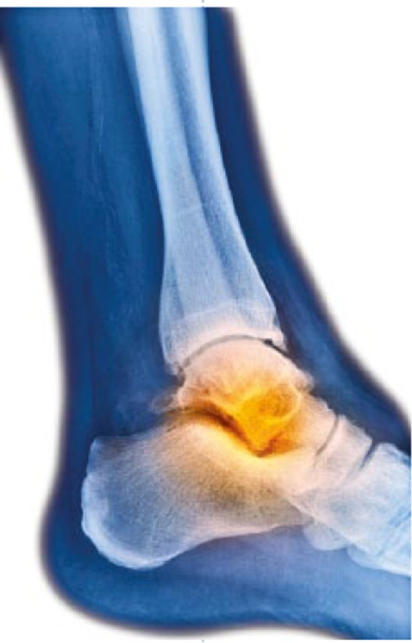
Fig.
Ankle and tibia the ‘D type’ fracture X-ref
The optimum treatment of tibial fractures with a synchronous ankle injury remains an unanswered question. Given the variety of fracture patterns in the tibia, and the variety of associated ankle fractures (e.g. some fibula alone, some with medial malleolar components), this is a question that is likely to be difficult to answer and may always be one of ‘expert opinion’. However, the occult ankle fracture associated with a tibial shaft fracture is a different beast. Surgeons in Cheonan (South Korea) raise the question of surveillance for the occult ankle fracture by describing the incidence and pattern of occult and overt ankle fractures in 77 consecutive patients with tibial fractures.4 The study team undertook CT scanning of the ankles of all patients and collated mechanistic, demographic and fracture pattern information to further describe the injury. Surprisingly, ankle fractures were visible on CT scanning in 64.7% (n = 47). Perhaps predictably, these authors identify the spiral tibial fracture as the morphology most likely associated with an occult posterior or medial malleolar fracture (89% incidence). These authors have a polarised view and come down strongly on the side of CT scanning all patients with tibial fractures. However, we do wonder if the presence of an occult fracture actually makes any difference to management? If it does not, then obtaining a CT scan just exposes the patient to needless ionising radiation.
Complications of frame treatment for Charcot foot deformity
Correction of deformity in the Charcot foot is often fraught with problems, but can be essential in limb preservation to prevent the accumulation of high plantar pressures resulting in skin breakdown. Jim Brodsky and others have, of late, popularised the use of frames to correct these complex and often resistant deformities, which has raised the obvious concerns of pin site complications and subsequent deep infection. This series from Loyola (USA) reports the outcomes of 283 patients, all with Charcot deformity treated with frame-assisted correction over an 11-year period.5 All surgery was achieved using a single-stage correction, and a 20% overall pin tract infection rate was reported. This is not dissimilar to complications of frame surgery in non-diabetics, and all settled with oral antibiotics with no requirement for wire removal. This series lends support and reassurance to surgeons undertaking frame correction of the Charcot foot. The authors highlight the importance of removing skin tension around wires, avoiding the use of half pins and constructs which allow the foot to trampoline on wires. The paper, however, contains no information on outcome or patient satisfaction with this method of correction; that is another question entirely.
Peroneal tendon instability in calcaneal fractures X-ref
Peroneal tendon pathology after calcaneal fracture surgery is a significant source of morbidity, and is often unrecognised. Subfibular impingement of the lateral wall blow-out is a well recognised indication for operative intervention, however, instability itself may remain unrecognised at a later stage. Pathology of the tendon system and the need to intervene acutely is a somewhat more controversial area. In an interesting collaboration from Rochester (USA), authors including Roy Saunders present an outcome series study of 155 calcaneal fractures with the aim of assessing the predictive value of tendon displacement on coronal CT as an indication of true disruption of the superior peroneal retinaculum (SPR) assessed at operation.6 Crucially, these authors have demonstrated that correlation is poor at best, and that this is probably due to the dynamic nature of the pathology. This is a very clinically useful paper as, on the one hand, it highlights the need to address the peroneal tendons, which changes the operative approach, and an extensile limb to the lateral incision is often needed. On the other hand, it highlights that surgery should not be decided for or against based on the CT findings of the peroneal tendons.
Early weight-bearing following subtalar arthrodesis
Isolated subtalar arthrodesis is a common salvage option and can provide reliable pain relief to patients with pain arising from the subtalar joint. While practice among surgeons differs, the majority prefer to limit early weight-bearing to promote union. However, in some patients the benefit of full loading of the affected limb is self-evident, allowing comorbid or non-compliant patients to achieve a reasonable result with minimal complications. There is rightly concern that this may promote fibrous nonunion at the prepared joint surface due to the torque stresses naturally occurring at the joint which is a product of its morphology, and maximising stability following fusion is a central surgical aim. A team from Innsbruck (Austria) has reproduced a cyclical loading model using a cadaveric specimen to perform biomechanical studies of differing screw stabilisation methods.7 Using eight paired subtalar joint specimens subjected to 1000 cycles at lower than physiological loads, the researchers tested both an angulated and parallel screw configuration for stability. They have demonstrated that the classic parallel screw technique yields significantly higher motion on cyclical testing than that measured at an interface bridged by two angled screws. The angled screw technique involves the standard inferior to superior screw with a start point on the posterolateral calcaneal tuberosity, but in addition includes a further, shorter screw capturing the anterior facet into the head of the talus. Even though only two possible configurations were considered, this does give some guidance for the surgeon who needs (often due to patient-centred factors) to allow a patient to weight-bear slightly earlier than might be ideal.
The tricky arthroscopic ankle arthrodesis: is it feasible to sneak in the back?
Arthroscopically assisted ankle arthrodesis offers many benefits, one being that in patients with poor soft tissues or profound diabetes, the soft-tissue complications are much less common than with open approaches. On occasion, however, a patient will present requiring an ankle fusion but the anterior soft-tissue envelope is not suitable for surgical wounds, even arthroscopically. The posterior ankle portal is rarely used in routine ankle arthroscopy due to perceived technical difficulty and the risk of iatrogenic injury to neurovascular and tendinous structures. The group from CHU in Rouen (France) present a feasibility study for arthroscopic ankle arthrodesis using an entirely posterior approach to the joint.8 Ten cadaveric specimens underwent fusion and were dissected afterwards to look for injury to structures around the ankle. None is reported in this small series, with an average operative time of 45 minutes. Operating in the real-life setting will inevitably be more time-consuming, but this study nicely demonstrates the feasibility of this method, to prepare the ankle via the posterior approach if needs be.
References
1. Agrawal Y , BajajSK, FlowersMJ. Scarf-Akin osteotomy for hallux valgus in juvenile and adolescent patients. J Pediatr Orthop B2015;24:535-540.CrossrefPubMed Google Scholar
2. Kramer DE , GlotzbeckerMP, ShoreBJet al.. Results of surgical management of osteochondritis dissecans of the ankle in the pediatric and adolescent population. J Pediatr Orthop2015;35:725-733.CrossrefPubMed Google Scholar
3. Adams SB , SettonLA, BellRDet al.. Inflammatory cytokines and matrix metalloproteinases in the synovial fluid after intra-articular ankle fracture. Foot Ankle Int2015;36:1264-1271.CrossrefPubMed Google Scholar
4. Jung KJ , ChungCY, ParkMSet al.. Concomitant ankle injuries associated with tibial shaft fractures. Foot Ankle Int2015;36:1209-1214.CrossrefPubMed Google Scholar
5. Ketz JP , MaceroliM, ShieldsE, SandersRW. Peroneal tendon instability in intra-articular calcaneus fractures: a retrospective comparative study and a new surgical technique. J Orthop Trauma2015. (Epub ahead of print)CrossrefPubMed Google Scholar
6. Finkler ES , KasiaC, KroinEet al.. Pin tract infection following correction of Charcot foot with static circular fixation. Foot Ankle Int2015;36:1310-1315.CrossrefPubMed Google Scholar
7. Eichinger M , SchmölzW, BrunnerA, MayrR, BölderlA. Subtalar arthrodesis stabilisation with screws in an angulated configuration is superior to the parallel disposition: a biomechanical study. Int Orthop2015;39:2275-2280.CrossrefPubMed Google Scholar
8. Malekpour L , RahaliS, DuparcF, DujardinF, RoussignolX. Anatomic feasibility study of posterior arthroscopic tibiotalar arthrodesis. Foot Ankle Int2015;36:1229-1234.CrossrefPubMed Google Scholar
