


Abstract
Arthroscopy has become a routine surgical procedure, used as a diagnostic and therapeutic tool for the treatment of joint problems. This article discusses its origins and looks at how it is currently used.
The name arthroscopy originated as a derivative of the Greek words 'arthron', meaning joint, and 'skopein', meaning look at. Perhaps one of the greatest revolutions in orthopaedic surgical care, arthroscopy has applications in every joint of the human body and is currently one of the most commonly performed orthopaedic procedures. It was first developed in the 19th century with the use of the cystoscope in cadaveric knees, and with subsequently more sophisticated dedicated arthroscopic equipment, it has become an invaluable component of modern orthopaedic care.
Historically, man's curiosity has always led to innovation and exploration. The human body is no exception, with much of the development of modern medicine and surgery attributable in some way to the urge to explore the 'inside' of the body; starting with the most basic, looking down throats and peering up rectums. This curiosity has been documented all the way back to the Roman Empire. Cavities and the inside of the human body, however, were not only of interest to the medical profession. It was known that artists during the Renaissance, such as Michelangelo and Da Vinci, had an interest in anatomical exploration, even under the threat of the Catholic Church.
Closed cavities, such as joints, were a specific challenge. It was easy to explore cadavers, but only the introduction of light and endoscopy allowed the exploration of a live joint. Endoscopic history goes, quite surprisingly, as far back as 1806, when Philipp Bozzini (1773 to 1809) (Fig 1) used an instrument called a 'lichtleiter' (Fig 2) which was designed to look into the bladder – the first endoscopic prodedure. The beginning of the endoscopic era was heralded by the development of the 'gazogene cystoscope' (Fig 3). This dangerous instrument, developed by Desormaux, provided light based on the combustion of gasoline and turpentine. Light was reflected into the bladder by a mirror.1
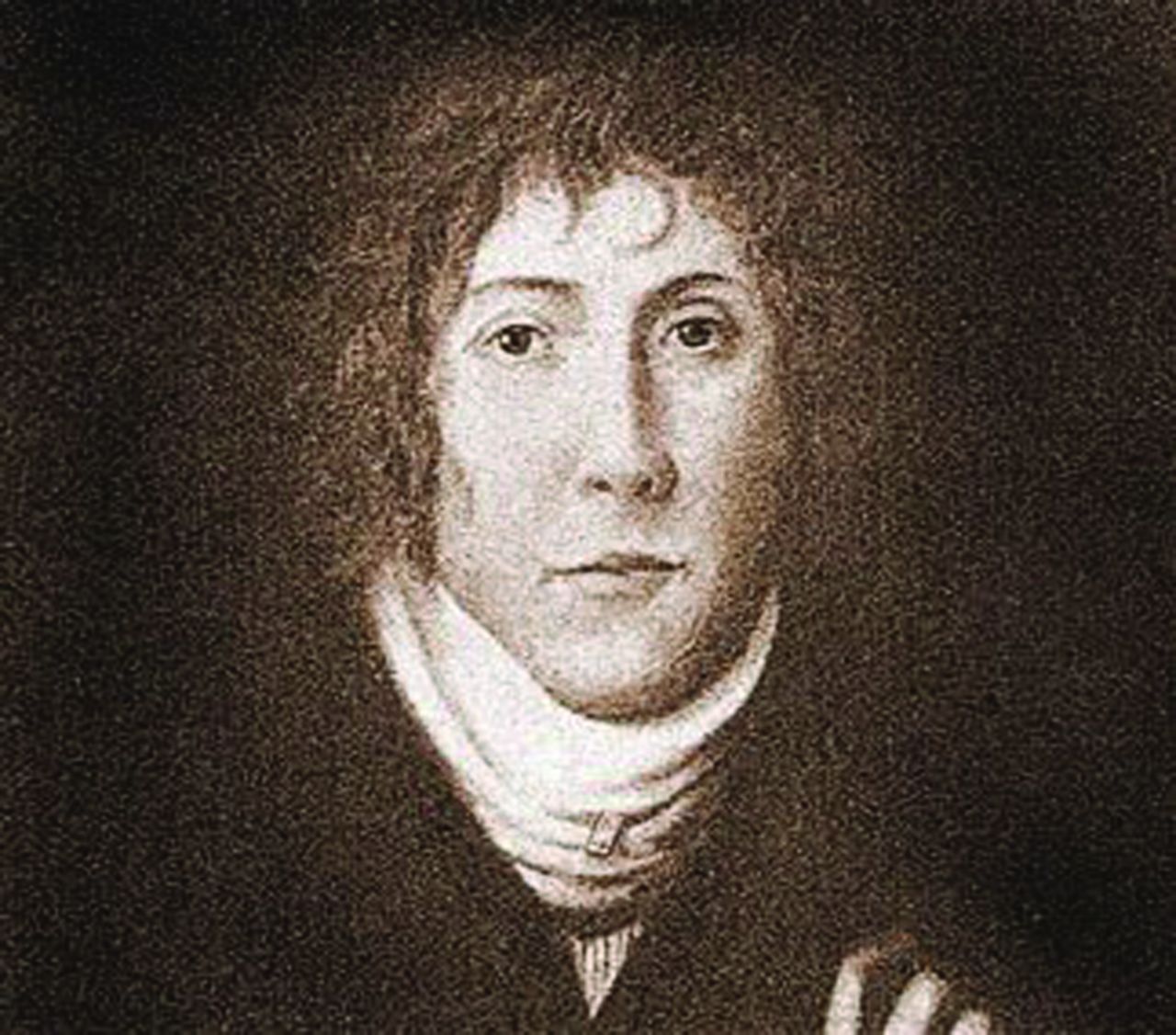
Fig. 1
Portrait of Bozzini.
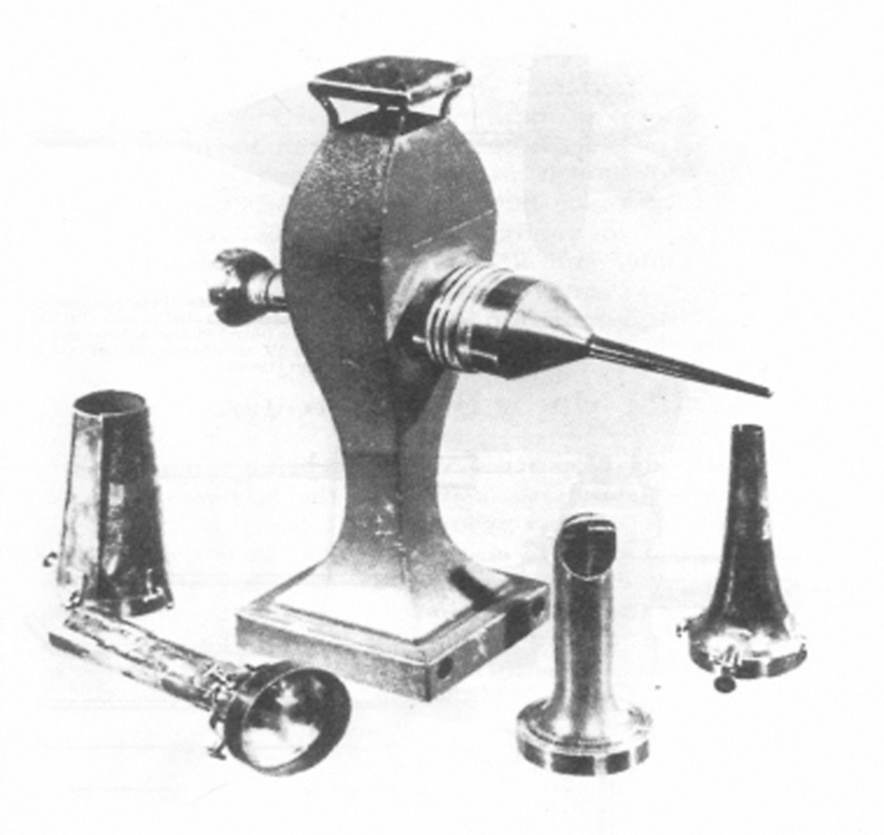
Fig. 2
Illustraition of a lichtleiter.

Fig. 3
Photograph of a gazogene cystoscope.
Thomas Edison's lightbulb, developed in 1879, was the next significant advance in endoscopy. A few years later, Leiter and Nitze developed the first incandescent bulb illuminating cystoscope,1 and the expansion of endoscopic exploration became inevitable. In Sweden, Hans Christian Jacobaeus (1879 to 1937) (Fig 4) started exploring the abdomen, and later the thoracic cavity, for the treatment of tuberculosis-related pleural adhesions, with the use of the 'laparo-thoracoscope'.2

Fig. 4
Photograph of Hans Christian Jacobaeus (1879 to 1937) at work.
Danish surgeon Severin Nordentoft (1866 to 1922) was the first individual to apply endoscopic instrumentation to a knee joint. The instrument he developed was a trocar of 5 mm in diameter, reportedly used in suprapubic cystoscopy, laparoscopy and knee arthroscopy. His report at the German Society of Surgeons in Berlin, 1912, was the first manuscript where the term 'arthroscopy' was used.3
In 1918, Kenji Takagi (1888 to 1963) (Fig. 5) used the cystoscope to visualise the cadaveric knee joint. He was the first true innovator and developer of arthroscopy. At that time in Japan, as in the rest of the world, tuberculosis was a very prevalent and serious disease. Takagi believed that early diagnosis of joint tuberculosis might lead to a better outcome following treatment. The concept of arthroscopy as an early diagnostic tool has not changed since.4

Fig. 5
Photograph of Kenji Takagi (1888 to 1963).
Like many other scientific advances, it is fascinating to see how similar concepts were developing independently throughout the world. In Switzerland, Dr Eugen Bircher (1882 to 1956) described the use of the arthroscope as an actual diagnostic tool in the treatment of meniscal pathologies. He would follow diagnostic 'arthroendoscopy', as he called it, with arthrotomy and appropriate surgery concurrently.5
Development of the arthroscope in the USA can be attributed to Phillip Kreuscher (1883 to 1943), an American surgeon born to German immigrant parents. His interest may have been thanks to his German background and time spent training at the Heidelberg University in 1912, or he may well have attended Nordentoft's presentation of knee endoscopy, as he was in Germany at the same time. Kreuscher returned to the USA, where he developed an interest in the treatment of injuries of the knee meniscus, resulting in an academic contribution and plea for early arthroscopy in athletic knee injuries.6 Michael Burman (1896 to 1974) (Fig. 6) was the next US surgeon to push forward the development of arthroscopic techniques. Burman continued the development of arthroscopic instrumentation and is credited with the development of a 4 mm diameter endoscope, as well as employing fluid for distension and visualisation of the joint. He is widely regarded as the first surgeon to attempt visualisation of the hip, and some of his published observations still hold true today.7

Fig. 6
Photograph of Michael Burman (1896 to 1974).

Fig. 7
Photograph of Robert Jackson.
In Japan, following the work of his mentor Dr Takagi and combined with the explosion of the electronic and optical industry, Masaki Watanabe (1911 to 1994) continued development of the arthroscope, culminating in the first colour photographs of the interior of the knee joint. He presented the first colour video at the SICOT Congress in Barcelona in 1957.8 Following this event, a Canadian, Robert Jackson (Fig. 7), showed an increased interest in the arthroscopic technique, and was able to arrange to work directly with Dr Watanabe in Japan. On his return to Toronto in 1965, the development of arthroscopy in North America started in earnest. Dr Jackson is a true founding father of the modern arthroscopy in North America, matching his own clinical interest with a shared professional interest, he was the founder of the International Arthroscopy Association as well as the Arthroscopy Association of North America.9
After 1965, arthroscopy as an orthopaedic surgical technique was starting to make its mark on the global orthopaedic community. Harold R. Eikalaar was the first person to achieve a PhD degree in arthroscopy, from the University of Groningen in the Netherlands. In Sweden, Ejnar Eriksson was a pioneer and early educator in arthroscopy, and the founding father of the European Society of Sports Traumatology, Knee Surgery and Arthroscopy (ESSKA), and Jan Gillquist became the pioneer of the central knee approach. From the United Kingdom, David J. Dandy, was an excellent teacher and was a co-author of the first text book on arthroscopy. David Marshall from Australia, together with David Dandy from the UK and Peter Fowler from Canada, established ISAKOS.
Although, historically, the time frame of the development of arthroscopy is short, the impact of the concept of minimally invasive surgery is enormous. There is currently no joint or cavity that can avoid the attention of the orthopaedic arthroscopist.
Shoulder
After the knee, shoulder arthroscopy was the next logical step because it used the same principles and technology applied to the knee, and often the same surgeons; in 1931, Michael Burman used the shoulder joint as part of his cadaveric work. The first clinical reports are the contribution of Andrén and Lundberg in 1965, describing a rigid shoulder treatment.10 Arthroscopy of the shoulder evolved more slowly than in the knee with more cautious early adopters. Conti in 197911 and Wiley in 198012 first presented their clinical experience of arthroscopic shoulder treatment.
Hip
Due to the anatomical differences and relative inaccessibility of the hip joint, the use of arthroscopy in the treatment of the hip has not followed the same pattern as the shoulder and is still struggling to gain acceptance in many institutions. Knee and shoulder arthroscopy developed from existing open procedures as the logical next step of tissue preservation, while hip arthroscopy was a completely new procedure. The presence of pathology and symptoms in the hip joint are traditionally treated by either conservative methods or radical replacement surgery. Arthroscopy of the hip joint offers both a logical alternative to what seems to be a commonly ignored condition, and a treatment conundrum; as a radically new treatment (now widely accepted), the precise indications, outcomes and contra-indications are still very much being debated.
The first attempt at surgical arthroscopy of the hip is again widely attributed to Dr Burman13 when, in 1931, he explored what would be called today a peripheral compartment, not using distraction. The next clinical application of hip arthroscopy is reported to have been by Dr Takagi in 1939, who used the technique on four patients.4 Following these early attempts, hip arthroscopy went unmentioned until the 1970s. In 1976, at the International Arthroscopy Association meeting, Aignan14 reported a large series of 51 diagnostic arthroscopies, reigniting interest in hip arthroscopy, which then transitioned from slow development to a gallop. Mirroring the rapid global uptake and interest in knee arthroscopy, the curiosity of surgeons across the world led to rapid developments in operative techniques and surgical instruments. Svante Holgersson and Ejnar Eriksson in Sweden, Richard Gross and James Glick in the USA, and Masaki Watanabe in Japan, were all pioneers in the heady days of early hip arthroscopy in the 1970s and 1980s, and interest in the procedure has not abated since. Thomas Sampson and John McCarthy contributed the lateral decubitus position, while in the UK Richard Villar played an essential role in the development of the procedure. Thierry Boyer and Henry Dorfmann in France, Michael Dienst in Germany and Hassan Sadri in Switzerland all developed their own variations on the techniques. Today hip arthroscopy has widespread acceptance, and the field of 'early intervention' is starting to offer the tantalising prospect of slowing or abating the progress of degenerative change in the hip joint.
Wrist
The development of small joint arthroscopy was inevitable after the successful application of arthroscopic techniques to other joints. Wrist arthrography was well established early in the 1980s as the gold standard for the detection of all varieties of wrist pathology including interosseous ligament injury. However, the detection and treatment of a wide range of wrist pathologies with an arthroscope had a major advantage due to the ability to visualise structures dynamically, and quickly gained favour with early adopters. The indications for wrist arthroscopy have continued to expand since Whipple's 1986 description of the new technique.15
Elbow
It is fascinating how the same people were involved in the pioneering of arthroscopy of all joints. The first mention of elbow arthroscopy was by Michael Burman in 1931. He reported a case of cadaveric elbow arthroscopy using a 4 mm endoscope and concluded that the elbow was 'unsuitable for examination'.13 Subsequently, development of the 1.7 mm endoscope by Watanabe in 1971, made the procedure suitable for small joints.8 Standardised approaches to elbow arthroscopy have only recently emerged due to the number of neurovascular structures crossing the elbow, with the newer approaches described only recently, in 1989 and 1990.16
Ankle
Ankle arthroscopy is a relatively recent development and certainly not yet a universally adopted technique. As with the wrist and elbow, smaller instruments for the restricted joint space needed to be developed. Nevertheless, Burman in 1931 scoped three cadaveric ankles using a 4 mm scope with no distraction. He stated: the ankle joint is not suitable for arthroscopic evaluation.13 Takagi was the first to perform arthroscopic ankle surgery, using the 2.7 mm instrumentation he developed for small joints. Watanabe took matters further, developing a 1.7 mm arthroscope, and describing the standard portals.4,17 By 1988, when Yates first described a non-invasive distraction technique18, ankle arthroscopy had developed into its current form with the development of the 2.5 mm, non-invasive distraction and irrigation system. Despite a relatively long history, like many more recent joints to undergo the attention of arthroscopists, there is still much debate as to which procedures are best performed arthroscopically and which open.
Spine
As technological development continues and arthroscopy proves to be safe and beneficial, early adopters are starting to evaluate arthroscopic surgery of the spine - perhaps the next challenge. The concept of minimally invasive spine surgery (MISS) was formed in the 1970s to address the long-term complication of extensive exposure of the surgical site and disregard of the integrity of the normal anatomical structures during spinal surgery. The contributions of Lyman Smith, Professor Hijikata, Professor Schreiber and Professor Leu in the field of MISS deserve recognition19 and paved the way for arthroscopic spinal surgery. Trans-thoracic thoracoscopic surgery is becoming commonplace and there are some early reports of minimally invasive posterior surgery. Time will tell if the innovations and revolution brought by the arthroscope to other orthopaedic disciplines do the same to the spine.
Conclusion
Arthroscopy began life as a curiosity and a desire to explore closed cavities and has developed into an essential part of modern orthopaedic surgery. Arthroscopic surgery has been a complete revolution to orthopaedic practice. Continual technological development has allowed the application of increasingly diverse arthroscopic techniques. Arthroscopy ranks amongst the most important contributions to orthopaedic surgery, and surgery in general, during the last century. Indispensable surgical skills, combined with exciting developments including genetic tissue engineering, laser treatment and robotics, mark arthroscopy out as one of the most continually evolving surgical techniques.
1 Jackson RW . A history of arthroscopy. Arthroscopy2010;26:91–103.CrossrefPubMed Google Scholar
2 Jacobaeus HC. über laparo- und thorakoskopie. In: Bauer L. Beiträage zur klinik der tuberkulose und spezifischen tuberkuloseforschung. Würzburg: Kabitzsch, 1912. Google Scholar
3 Nordentoft S. Über endoskopie geslossener cavitaten mittels eines trokar-endoskopes. Verh Dtsch Ges Chir 1912;41:78-81. Google Scholar
4 Takagi K . The arthroscope: the second report. J Jpn Orthop Assoc1939;14:441–466. Google Scholar
5 Kieser CW , JacksonRW. Eugen Bircher (1882-1956) the first knee surgeon to use diagnostic arthroscopy. Arthroscopy2003;19:771–776.CrossrefPubMed Google Scholar
6 Kreuscher P . Semilunar cartilage disease: a plea for the early recognition by means of the arthroscope and early treatment of this condition. Ill Med J1925;47:290–292. Google Scholar
7 Byrd JWT. Operative hip arthroscopy. Second ed. Springer: New York, 2005. Google Scholar
8 Watanabe M . Memories of the early days of arthroscopy. Arthroscopy1986;2:209–214.CrossrefPubMed Google Scholar
9 Jackson RW . Memories of the early days of arthroscopy: 1965-1975: the formative years. Arthroscopy1987;3:1–3. Google Scholar
10 Andrén L , LundbergBJ. Treatment of rigid shoulder by joint distension during arthrography. Acta Orthop Scand1965;36;45-53.:. Google Scholar
11 Conti V . Arthroscopy in rehabilitation. Orthop Clin North Am1979;10:709–711. Google Scholar
12 Wiley AM , OlderMW. Shoulder arthroscopy: investigation with a fiberoptic instrument. Am J Sports Med1980;8:31–38. Google Scholar
13 Burman MS . Arthroscopy or the direct visualization of joints: an experimental cadaver study: 1931. Clin Orthop Relat Res2001;390:5–9. Google Scholar
14 Aignan M . Arthroscopy of the hip. Rev Int Rheumatol1976;33:458. Google Scholar
15 Whipple TL , MarottaJJ, Powell JH 3rd. Techniques of wrist arthroscopy. Arthroscopy1986;2:244–253.PubMed Google Scholar
16 McGinty JB, Burkhart SS, Jackson RW, et al. Operative arthroscopy. Third ed. Philadelphia: Lippincott Williams & Wilkins, 2003. Google Scholar
17 Takagi K. The arthroscope. J Jpn Orthop Assoc 1939;14:359-441. Google Scholar
18 Yates CK , GranaWA. A simple distraction technique for ankle arthroscopy. Arthroscopy1988;4:103–105.CrossrefPubMed Google Scholar
19 Kambin P . History and evolution of arthroscopic & endoscopic lumbar disc surgery: the U.S. Experience. Internet J Minim Invasive Spinal Tech2007;1.:. Google Scholar
