


Abstract
Ancient Egypt was a highly developed agrarian society with a massive civil engineering capability. Trauma and skeletal disease were common and vestiges of the evidence for that survive, largely in the form of hieratic images and papyri dedicated to the practice of medicine. The earliest treatise on trauma is the Edwin Smith papyrus, possibly the work of Imhotep. This study details some remarkable examples of musculoskeletal pathology including fatal open fractures, foot deformity of Tutankhamun, and the earliest recorded instances of child abuse.
Ancient Egypt was a thriving and bustling civilisation. Massive civil engineering projects, war and disease brought a great variety of medical conditions on the Egyptians. The disease burdens of trauma, as well as orthopaedic conditions, are well-evidenced. Yet the musculoskeletal care of those times was far from primitive. Indeed, a proportion of what we now regard as up-to-date orthopaedic and trauma care can trace its roots back to ancient Egypt.
Ancient medical texts
Perhaps the most valuable text on medicine from this period of the ancient world is the so-called Edwin Smith Papyrus. Other medical papyri exist, such as the Ebers Papyrus and London Medical Papyrus, but they are largely rooted in magic. The Edwin Smith Papyrus only refers to metaphysical spells only twice, and is almost entirely analytical. The papyrus dates from the 17th century BC, and is arguably the earliest known surgical treatise. The manuscript was so extensive that it runs to more than 15 feet in length.
The manuscript was acquired in Luxor in 1862 by the American Edwin Smith, a pioneer Egyptologist, who bought it from an Egyptian dealer named Mustafa Agha. After Smith’s death in 1906, the papyrus was donated by his daughter to the New York Historical Society, allowing James Henry Breasted to study it from 1920 onwards. A translation, transliteration and discussion were published by Breasted in 1930.1
Breasted argues that the Edwin Smith Papyrus is in fact a later copy of an ancient manuscript, containing the original author's text from 3000 to 2500 BC, combined with a later compendium of 69 explanatory notes. The authorship remains a matter of debate, some even attributing it to Imhotep (circa 2650 to 2600 BC), an architect, high priest, and physician of the Old Kingdom.
The papyrus consists of systematically arranged accounts of 48 trauma cases, rather than instructional dicta, starting with the head and proceeding caudad, but below the thorax and spine is absent.
The cases addressed are:
27 head injuries (cases 1 to 27)
6 throat and neck injuries (cases 28 to 33)
2 injuries to the clavicle (cases 34 to 35)
3 injuries to the arm (cases 36 to 38)
8 injuries to the sternum and ribs (cases 39 to 46)
1 injury to the shoulder (case 47)
1 injury to the spine (case 48)
Each case is classified by one of three different verdicts: 1) ‘An ailment that I will treat’; 2) ‘An ailment with which I will contend’; or 3) ‘An ailment not to be treated’. The latter comprises those cases regarded as incurable, such as a case of cervical spinal injury with quadriplegia (Case 31), and another of a depressed temporal bone fracture with aphasia (Case 8). To this extent, the Edwin Smith papyrus represents the origin, and earliest record of medical triage.
One of the most clearly described cases (Case 35) details management of a patient with a fracture of the clavicle (Fig. 1):
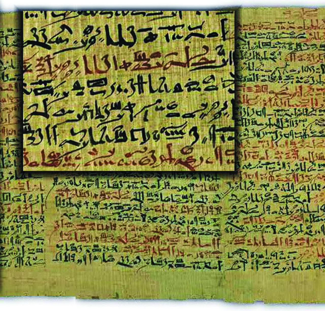
Fig. 1
Section of Edwin Smith papyrus describing a clavicular fracture.
Instructions concerning a break in his collar bone. If thou examinest a man having a break in his collar-bone and thou shouldst find his collar-bone short and separated from its fellow, thou shouldst say concerning him, 'One having a break in his collar-bone, an ailment which I will treat.'
Thou shouldst place him prostrate on his back, with something folded between his two shoulder-blades; thou shouldst spread out his two shoulders in order to stretch apart his collar-bone until that break falls into its place, thou shouldst make for him two splints of linen, and thou shouldst apply one of them on the inside of his upper arm and the other below his upper arm. Thou shouldst bind it with ymrw, and treat it afterward with honey every day, until he recovers.
The earliest records of human fracture splintage
An expedition, led by Dr George Reisner between 1903 and 1904, and funded by Mrs Phoebe Hearst, wife of the American press magnate, William Randolph Hearst, excavated a site at Naga ed Der, 100 miles north of Luxor. The archaeological finds of the dig included a number of human specimens with definite fractures, two of which had been treated with splintage.2 The splints unearthed in the excavation comprised a system of linen-wrapped, palm bark boards placed carefully over the limb with sufficient padding. These were held in place with linen bandages. The specimens clearly demonstrated well-evolved fracture splintage.
The femoral specimen (Fig. 2) is that of a female adolescent with a fracture of the femoral shaft. A wedge fragment of the bone is missing, and the palm fibre pad found adherent to the splint at the fracture site bore traces of blood pigment, indicating an open fracture. There is no evidence of any periosteal reaction and the victim is thought to have died early after injury. Such an open femoral fracture would have been unlikely to have been survivable at that time – presumably, the author of the Edwin Smith papyrus would have described this as ‘an ailment with which I will contend’ – worth a go at treating but with little expectation of a cure.
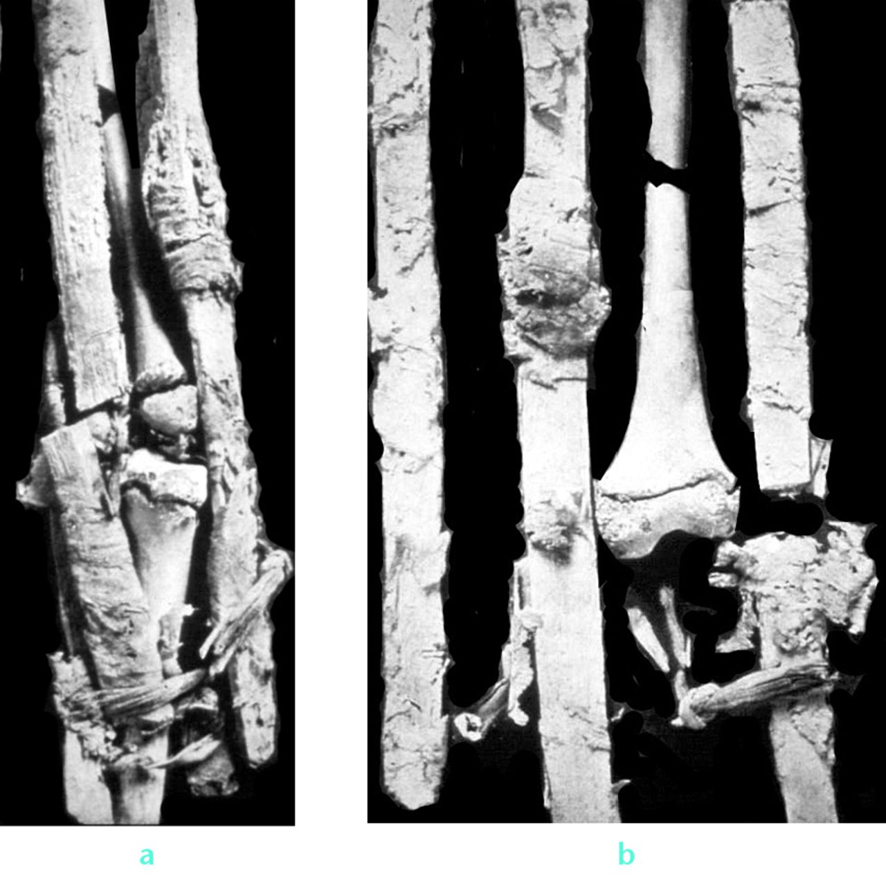
Fig. 2
Photographs (a and b) of the earliest known treated human fracture specimen: open femoral fracture of an adolescent (note open physes).
The second patient with evidence of fracture treatment had suffered a forearm injury with both bones fractured. In this case, the splint was made of stacked reeds and three roughly-shaped braces of palm bark. Blood-stained, date-palm fibre was found adherent to the end of the proximal ulnar fragment, probably having been inserted in order to arrest bleeding. Again, there were no signs of any significant callus formation and early death was also likely to have occurred.
In a society such as that of ancient Egypt in which fractures were common and some documentation of treatment methods has been found, it is surprising that there is scant evidence of any writing concerning joint dislocations. However, in an iconic painting in the tomb of Ipuy, circa 1300 BC, the sculptor of Ramses II, a funereal artist has depicted a physician reducing a dislocated shoulder, using a technique similar to that which Kocher described in 1870,3 some 3200 years later (Fig. 3). It is unknown how this relates to the life, or death, of Ipuy. It is entirely possible that a worker at the tomb sustained such an injury and the painter’s experience of witnessing the reduction manoeuvre inspired him to record the incident. This is the oldest known image of shoulder dislocation and of an attempt at a reduction.
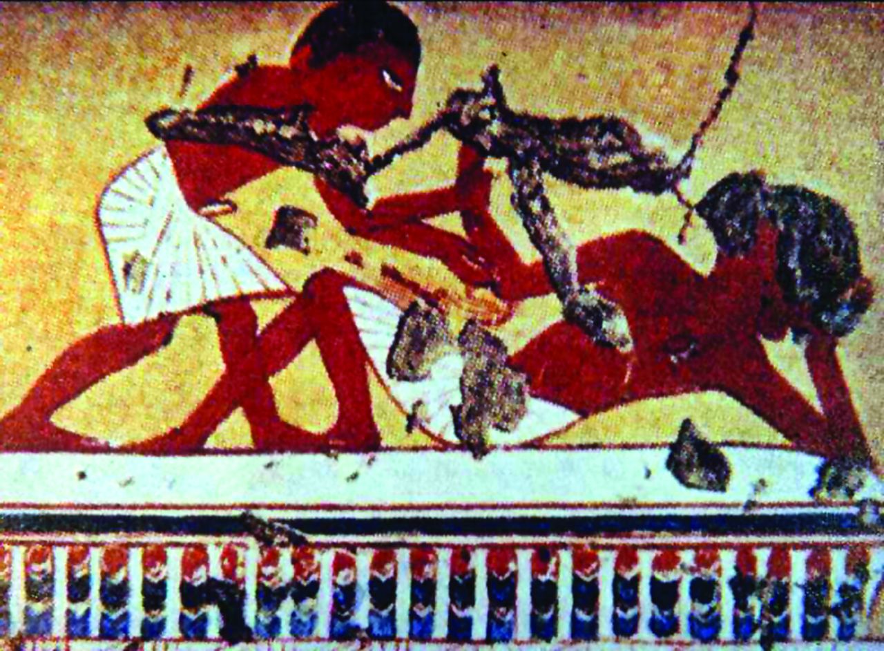
Fig. 3
Painting from the entrance to the tomb of Ipuy, circa 1300 BC.
Dislocation of the shoulder continued to be a severely disabling and common injury throughout early history. The records of antiquity reflect the seriousness of the treatment problem, as evidenced by the powerful machines described by Hippocrates (glossocomium) and later so beautifully illustrated by Vidus Vidius (1509 to 1569).4 The theme of shoulder dislocation in antiquity may in part be due to the often considerable delay in presentation.
Surgical fracture care
Surgical care of fractures is highly unlikely to have been practised so many millennia ago. There is however some mystery surrounding the sarcophagus of Usermontu. This 18th dynasty burial casket is preserved at the Rosicrucian Museum in San José, California. The casket was acquired in an amusing manner in 1971 having appeared in a Neiman-Marcus Christmas catalogue, in a section entitled ‘His and Her Gifts for People Who Have Everything’.
Usermontu was a vizier to Tutankhamun during the 18th Dynasty and was the son of Besenmut, priest of the god Montu, Lord of Thebes. The mummy within the sarcophagus is thought not to be that of Usermontu himself, but an unknown mummy substituted by ancient grave robbers. In fact, carbon-14 dating suggests that the mummy died around 600 BC, and not the 1300 BC date of the sarcophagus. Those ancient bodysnatchers actually did an unintentional great service to medical archaeologists: the mummy from Usermontu’s sarcophagus came under the scrutiny, in 1996, of a medical team spearheaded by the renowned Egyptologist Dr Wilfred Griggs. Dr Griggs is the director of Ancient Studies at Brigham Young University and the Professor of Ancient Scripture. The forensic medical examination uncovered what appeared to be evidence of previous surgery to the right knee (Fig. 4). Radiographs demonstrated an intramedullary fixation achieved with an iron nail (Fig. 5).Griggs came to the conclusion that the implant had been inserted around the time of death: ‘It didn't appear to be a modern pin insertion. There were some characteristics of the knee joint, namely some of the ancient fats and textiles that were still in place and would not have been in place had the leg been separated in modern times.’
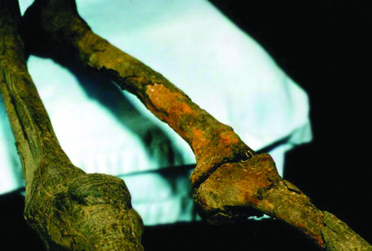
Fig. 4
Photograph of the right knee of the mummy of Usermontu.
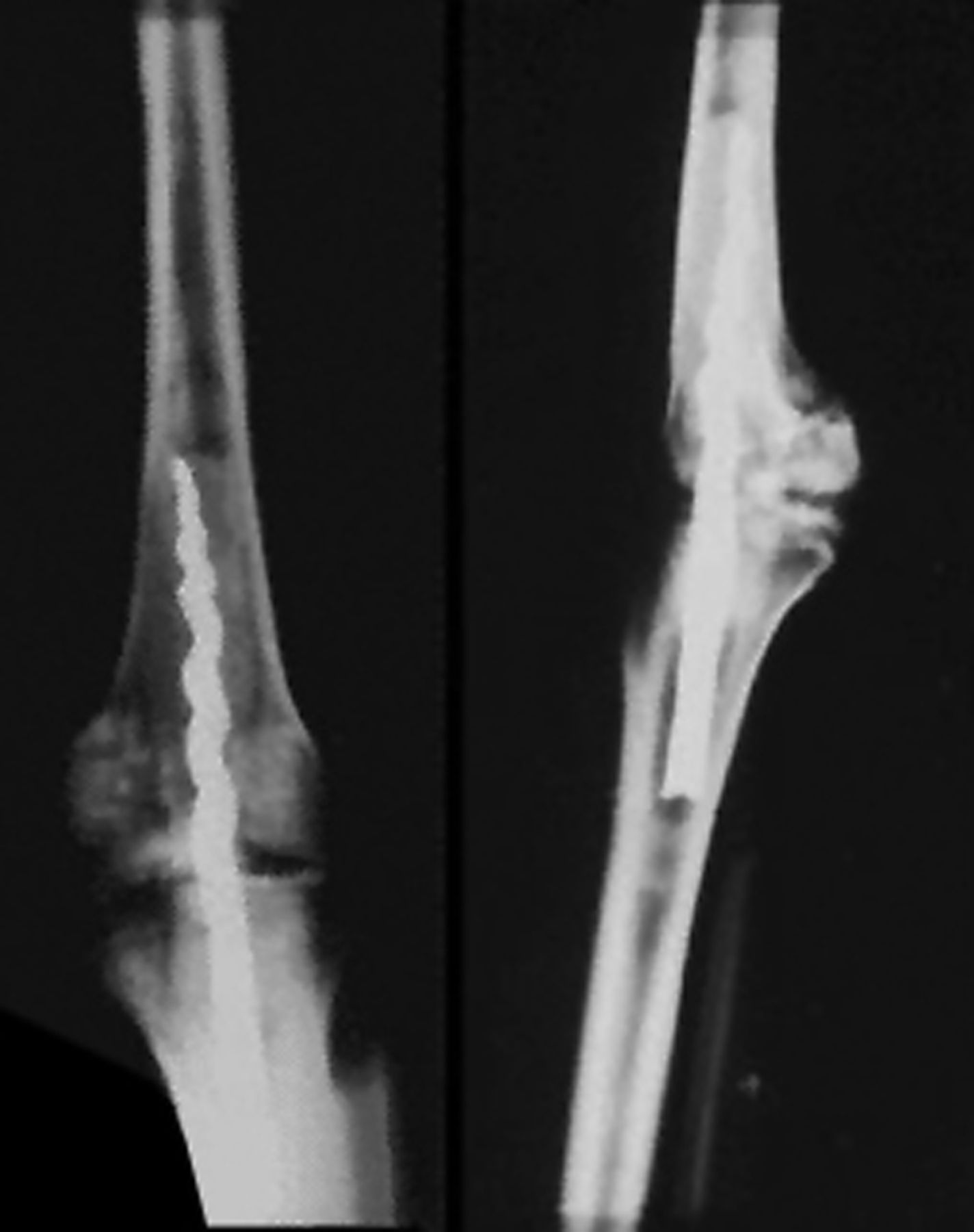
Fig. 5
Radiograph of the right knee of the mummy of Usermontu.
The pin, reported Griggs, tapers into a corkscrew as it enters the femur; the other end of the pin, positioned within the tibia, has three flanges (shades of Smith-Petersen) extending outwards from the core of the pin, thereby preventing rotation of the pin within the bone. The cavity for the pin in the tibia also contained a resinous, glue-like material that aided in the fixation of the joint. Samples of the resin revealed it to consist of a type of cedar gum, mixed with other organic materials.5
The Ancient Egyptians believed strongly in a physical resurrection and recombination with the soul in the afterlife. It is thought that the technicians embalming the cadaver, were preparing the body for its reunification with the spirit. It was observed that great surgical skill had been brought to bear during the insertion of this nail, suggesting that it could well have been a common practice, at least among embalmers.
King Tutankhamun, whom Usermontu served, ruled ancient Egypt from 1333 BC to 1324 BC, during the 18th dynasty (1550 to 1295 BC) of ancient Egypt's New Kingdom. His brief reign, youthful appearance, and premature death at the age of about 18 years, as well as the dramatic rediscovery of his remains and tomb in 1922 by Howard Carter, captured the imaginations of Egyptologists, historians and the public for decades. The manner of the young pharaoh’s death remains the subject of heated debate and voluminous literature, scientific and popular.6 DNA of Plasmodium vivax has been found in the mummy, indicating that he suffered from malaria. More interesting has been the finding of a fresh, supracondylar femoral fracture, with traces of embalming materials adherent to the bone surfaces, indicating a possible open fracture: such a fracture would have most likely been fatal at that time (see from examination of splinted fractures from Naga ed Dehr above). What is certain is that radiographs and CT scans of his feet show that he had a deformed left foot, right talocalcaneal synostosis (Fig. 6)and also osteochondritis of the left second metatarsal head (Fig. 7).7
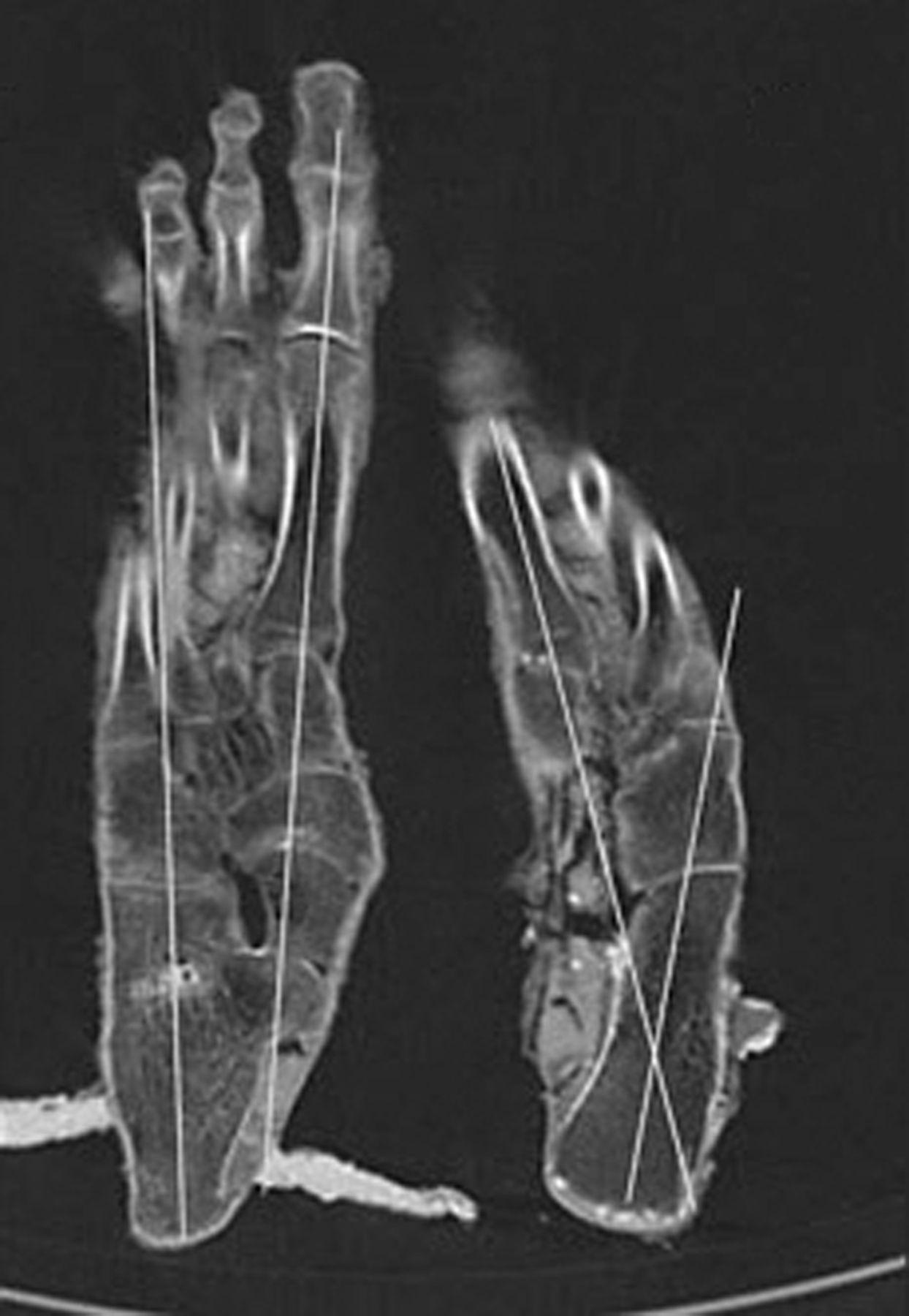
Fig. 6
CT scan of Tutankhamun’s feet.
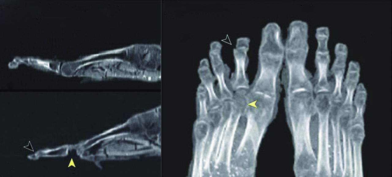
Fig. 7
Radiographs of Tutankhamun’s feet, showing osteochondritis of left 2nd metatarsal head.
Also found In Tutankhamun’s tomb was a collection of walking staffs, resplendent with beautifully carved heads (Fig. 8), all showing evidence of wear, and therefore use, at their tips. These are thought to have been the pharaoh’s own walking aids, and he is often depicted as walking, or standing, with the aid of a staff (Fig. 9).
Fig. 8
Beautifully decorated walking staffs from Tutankhamun’s tomb.
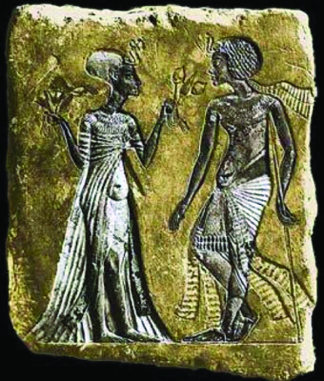
Fig. 9
Stele thought to be of Tutankhamun using a walking staff – note the short, thin left leg.
Perhaps one of the most disturbing aspects of modern society is the tragic prevalence of physical violence directed at children. The involvement of physicians in the care of victims of child abuse may not be limited to modern civilisations and cultures. In such a context, we see that this shameful attribute of human society has existed for millennia. There exists a fascinating court record, the Papyrus Oxyrynchus 6.903,8 which offers a graphic account of a man accused of abusing his young slaves and his wife’s foster-daughters, as follows:
Accusation against a husband. Concerning all the insults uttered by him against me. He shut up his own slaves and mine with my foster-daughters and his agent and son for seven whole days in his cellars, having insulted his slaves and my slave Zoe and half killed them with blows, and he applied fire to my foster-daughters, having stripped them quite naked, which is contrary to the laws. He also said to the same foster-daughters, ‘Give up all that is hers,’ and they said, ‘She has nothing with us’; and to the slaves when they were being beaten he said, ‘What did she take from my house?’ and they under torture said, ‘She has taken nothing of yours, but all your property is safe.’ Zoilus went to see him because he had shut up his foster-son, and he said to him, ‘Have you come on account of your foster-son, or of such a woman, to talk about her?’
The records of ancient Egyptian child abuse are not limited exclusively to papyri. A striking find was made, by Sandra Wheeler and colleagues of the Departments of Anthropology of the University of Western Ontario and the University of California, Berkeley,9 in the Kellis 2 Cemetery. The cemetery is a Roman Period (50 to 450 AD) burial place, located at the Dakhleh Oasis, Egypt. The investigative team reported some of the only archaeological evidence of a probable case of repeated malintent towards a young child, thereby giving the occurence of physical child abuse considerable time depth, as this may be the earliest documented case. The skeletal remains of this child (Fig. 10), aged between two and three years, exhibited fracture patterns consistent with chronic physical abuse. This seems likely to have led to the child’s untimely death and represents the earliest documented record of non-accidental injury.
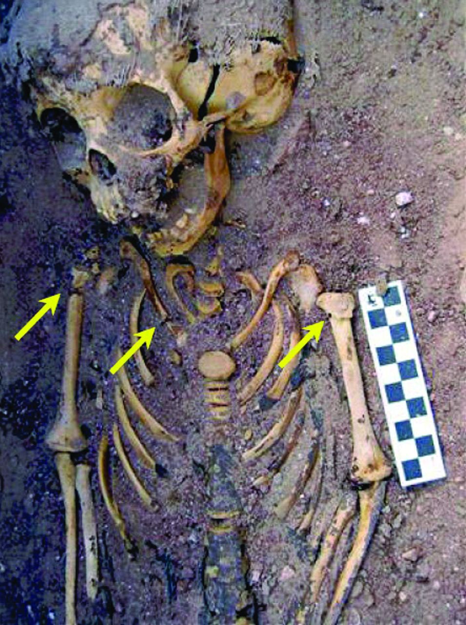
Fig. 10
Skeletal injuries in Kellis 2 specimen, strongly suggesting non-accidental injury.
Macroscopic, radiological, histological and isotopic analyses were used to determine if any physical effects of abuse were detectable in this child’s remains. Their results supported such a diagnosis.
The distribution of fractures and subperiosteal new bone formation (SPNBF) in this child are shown in Figure 11. Active cribra orbitalia (porotic hyperostosis of the orbital margins, usually associated with chronic stress or anaemia) were noted in both orbits, as well as slight SPNBF on the temporal and zygomatic processes, and on the medial and lateral surfaces of the mandibular condyles. The humeri exhibited complete fracture of the proximal third of each diaphyses. Early callus bridged across the fracture site on the left humerus, suggesting that the fracture occurred at least two weeks prior to death. Evidence of healing is also apparent on the margins of both fractures, as well as in trabecular bone within the proximal portion of the humeri. Periosteal reaction enveloped the diaphysis of the right radius, and distal portion of both ulnar diaphyses exhibited asymmetrical new bone formation. There was a complete fracture of the medial aspect of the right clavicle, with no evidence of healing, indicating a peri-mortem injury. Both scapulae bear evidence of SPNBF on the ventral and dorsal surfaces. Two well-healed fractures are visible in the 7th left and 8th right ribs with mature callus formation. Most ribs revealed periosteal reactions on the external and visceral surfaces, in varying degrees, most pronounced in the lower ribs. The right ilium showed plastic deformation on the superior aspect of the iliac wing.
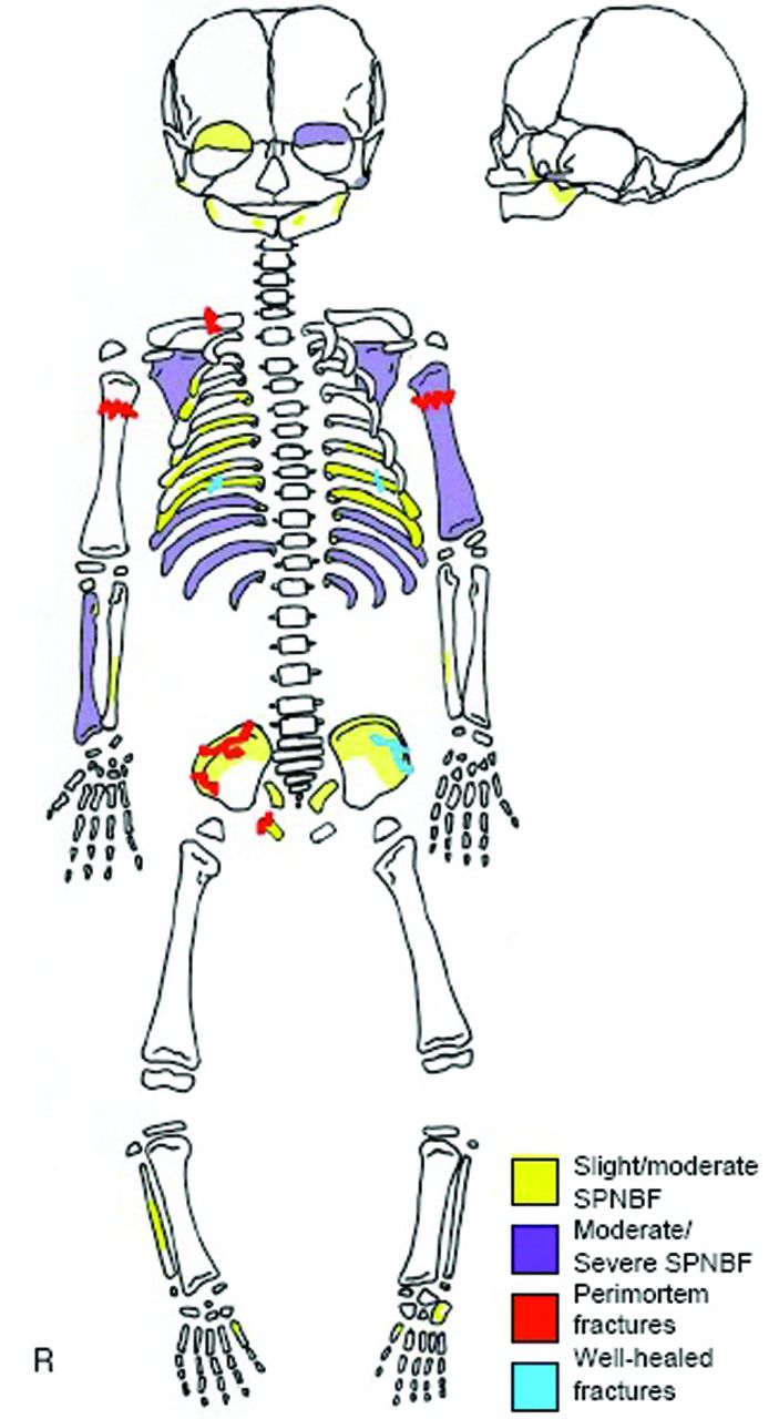
Fig. 11
Distribution of skeletal injuries in Kellis 2 specimen, strongly suggesting.
Discussion
There is a wealth of evidence, selections from which are presented here, that orthopaedic pathology, especially trauma and including non-accidental injury of children, faced the physicians of Ancient Egypt. What has been found hitherto can only represent a tiny fraction of the true picture and incidence of these problems in that long-lasting and very well-developed society.
Despite being highly developed in many aspects - architectural, artistic, gubernatorial and religious - Ancient Egypt lacked effective therapies for severe injury, including open fractures. Indeed, it was not until the 20th century that open fractures did not carry a probable, lethal risk. Billroth10 reported 93 patients with open fractures of the lower leg (1860 to 1887), of whom 39% died. Even in those treated with amputation 20 of 28 (71%) died. Of the 16 patients Bilroth reported with open femoral fractures, ten died, giving a mortality rate of 63%.
There is no accessible evidence that the Ancient Egyptian physicians practised amputation for open fractures: ablation of the nose as a judicial punishment was not uncommon and circumcision was widely performed, with several surviving pictorial depictions of the latter. It is likely that contemporary culture was restrained by taboos and religious imperatives to preserve bodily unity, particularly the desire to enter the afterlife ‘whole’, ensuring that elective amputation did not take place.
The Smith and Ebers Papyrus and other documents of ancient Egypt offer no account of surgical amputation.11 Nevertheless, traumatic amputation, disease and congenital abnormality resulting in loss of an appendicular part, probably led to the construction of prosthetic replacements.
During an excavation campaign at the necropolis of Thebes-West (built 1550 BC to 1300 BC), by the German Institute of Archaeology, Cairo, and the Supreme Council of Antiquities in Egypt, Professor A. Nehlich and colleagues discovered a fragmented, but otherwise well-preserved, mummy of a woman. Palaeopathological examination showed that the big toe of the right foot had been amputated. The toe had been removed during her lifetime, the amputation site being covered by an intact layer of soft tissue, including skin. The missing toe had been replaced by a wooden prosthesis (Fig. 12), painted dark brown and made up of three separate components. The main component consisted of a longitudinal wooden corpus that replaced the toe. This was attached to two small wooden plates, which were fixed to each other by leather thongs. All wooden parts were delicately manufactured and the corpus of the prosthesis perfectly shaped like a big toe, even including the nail. A broad textile strap was fixed to the small plates and to the prosthetic corpus, and tied around the forefoot, fixing the prosthesis firmly in place. This construction provided sufficient stability to keep the prosthesis in the correct position, and to allow the user to move without major restrictions. Careful inspection revealed clear marks of use on the plantar aspect of the toe.12 This appears to be the oldest known prosthesis. It is possible, however, that Egyptian embalmers used similar prostheses to restore physical completeness in people headed to the afterlife with mutilated feet, or limbs.
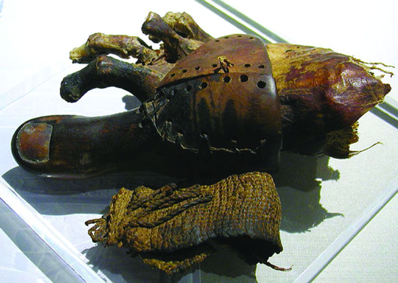
Fig. 12
Ancient Egyptian hallucial prosthesis, made of wood and leather.
From the available evidence, it seems that the practice of orthopaedic care was widespread and, given the constraints of the time, most inventive. Whereas, in the modern context, there may be few practically applicable lessons to be learned from study of this subject, we can be suitably humbled and therefore stripped of complacency, by being able to admire the industry and dedication of our professional forebears, practising our art over 3000 years ago. Let us never forget that triage of the wounded was first described by an unknown savant 5000 years before Baron Dominique-Jean Larrey.
1 Breasted JH. The Edwin Smith surgical papyrus. Chicago: University of Chicago Press, 1930. Google Scholar
2 Smith GE . The most ancient splints. Br Med J1908;1:732–736.CrossrefPubMed Google Scholar
3 Kocher T. Eine neue reductions methode fur schulter-verrenkung. Berlin; Klin, 1870 (in German). Google Scholar
4 No author listed. Vidus Vidius (1508-60). Med Hist 1957;1:186-188. Google Scholar
5 Snow K. BYU professor finds evidence of advanced surgery in ancient mummy. Brigham Young University Magazine, June 1996. http://magazine.byu.edu/?act=view& a=483 (date last accessed 4 March 2013). Google Scholar
6 Markel H . King Tutankhamun, modern medical science, and the expanding boundaries of historical inquiry. JAMA2010;303:667–668.CrossrefPubMed Google Scholar
7 Hawass Z , GadYZ, IsmailS, et al.Ancestry and pathology in King Tutankhamun’s family. JAMA2010;303:638–647. Google Scholar
8 No author listed. Papyrus Oxyrhynchus 6.903. The Speer Library, Princeton Theological Seminar, Princeton, New Jersey, USA. http://www.papyri.info/hgv/33342 (date last accessed 5 March 2013). Google Scholar
9 Wheeler SM, Beauchesne P, Williams L, Molto JE. Fractured Childhood: A Case of Probable Child abuse from Ancient Egypt. SAS Bulletin: Newsletter of the Society for Archaeological Sciences 2007;30:6-9. Google Scholar
10 Billroth T. Die allgemeine chirurcische Pathologie und Therapie in 50 Vorlesungen. Berlin:Verlag Georg Reimer, 1866 (in German). Google Scholar
11 Ghalioungui P. La Médecine des Pharaons. Paris: Laffont, 1983. Google Scholar
12 Nerlich AG , ZinkA, SzeimiesU, HagedornHG. Ancient Egyptian prosthesis of the big toe. Lancet2000;356:2176–2179.CrossrefPubMed Google Scholar
