X-ref For other Roundups in this issue that cross-reference with Children’s orthopaedics see: Foot & Ankle Roundups 1, 2; Hip Roundup 6; Oncology Roundups 4, 5; Spine Roundup 5.
The end of the ‘What’ test?
A commonplace explanation for trainees as to how to choose patients for prophylactic pinning of the contralateral slipped capital femoral epiphysis (SCFE) is the ‘What test’. When explaining to the family that the other side may also need surgery and to return immediately if the child limps or there is any discomfort, and the reply from the parents is “What?”, the pragmatic approach is prophylactic fixation. This approach, while common sense-based, presumes a relatively low contralateral slip rate and that all of the risk of surgery is related to the surgery and not to the anaesthetic. Funnily enough there aren’t very many studies evaluating the contralateral slip rate, and fewer still comparative outcome studies of prophylactic fixation versus a wait-and-see approach. The paediatric team in Edinburgh (UK) report their experience of 86 patients managed with a combination of strategies in a retrospective case series.1 The research team reports the outcomes of 54 males and 32 females, 50 of whom underwent unilateral fixation and 36 of whom underwent bilateral fixation. In their series the risk of subsequent slip was 46% in the unilateral group, and for comparison there were poorer quality of life scores and complication risks in the group who did not receive prophylactic pinning. The study team has calculated the cost per QALY to be £1431, well below the cost-effectiveness threshold for NICE recommendation in the UK. Given the results presented here, certainly in our practice here at 360, the ‘What test’ is out and prophylactic fixation is in.
Bone marrow implantation in osteonecrosis of the femoral head
Osteonecrosis of the femoral head is a catastrophic and disabling condition when it progresses to complete collapse and osteonecrosis. Although there is much research concerning Perthes’ disease, interventions and outcomes, there is surprisingly little on the commonest global cause of osteonecrosis – sickle cell disease. Researchers in Philadelphia (USA) report their early results of epiphyseal drilling and autologous bone marrow implantation.2 In a small series of 14 hips, the authors report the outcomes to a year following surgery based on clinical (Children’s Hospital Oakland Hip Evaluation Scale), radiographic signs of involvement and hip range of motion. While there was no comparator group (and as such, natural history of the disease was not taken into consideration), significant improvements were seen in pain and range of motion, however, there were no changes in progression of anterior and lateral collapse. This study suggests that this treatment may offer a surgical option to an otherwise difficult to treat group of patients. Without a comparator group and with the dual interventions it is difficult to know if the treatment effect is real (or just the natural history of the disease), or if the autologous bone marrow offers any treatment effect over drilling alone. A comparative study is clearly needed to draw any conclusions here.
Subphyseal Puddu correction X-ref
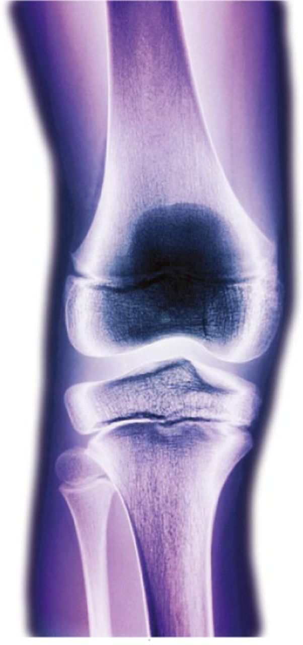
The Puddu technique of osteotomy with a wedged plate is a well-established and respected technique. While the track record is proven for this established technique, authors in Damanhur (Egypt) report their experiences with a modified subphyseal approach to the tried and tested Puddu technique.3 The authors report the outcomes of 25 legs, all treated for tibia vara in 18 adolescents. Outcomes were reported to one year and, using a technique that should afford greater options for angular deformity, the authors were able to achieve angular corrections of 22 degrees with no incidences of neurological deficit or interference in growth within the proximal physis.
Gunshot injuries in the child X-ref
Though thankfully not a problem in many parts of the world, this article from authors in Houston (USA) nevertheless provides a valuable insight into the problem of open fractures and gunshot-associated injuries in the child.4 Their paper is a retrospective review of children admitted with gunshot fractures in two level 1 major trauma centres, and concerns the outcomes of 49 children with 58 identified fractures. The most common sites of injury were the tibia and femur (40% of injuries), with associated injuries present in 47% (usually abdominal, genito-urinary and neurovascular injuries). In a centre with a clearly vast experience in these injuries, just over half (63%) underwent debridement in the operating theatre, with the remainder likely to have had ‘bedside debridement’. The overwhelming majority were managed non-operatively (71%) from the fracture perspective. The authors propose an algorithm based on four decisions and recommend that this is introduced into the management of this injury. There are perhaps no surprises about the demographics as these patients will involve combatants and innocent, predominantly male, bystanders. What is surprising is the lack of structure to the management in a US level 1 trauma centre and this paper goes some way to rationalising the treatment and provides a sensible algorithm which could be used in UK practice.
Scaphoid fractures in the child X-ref
Another tricky injury to rationalise based on research is the paediatric scaphoid fracture. Surgeons in Iowa City (USA) report the outcomes in 63 of a cohort of 312 patients with scaphoid fractures, aged between eight and 18 years at the time of treatment.5 Outcomes were assessed at a median follow-up of 6.3 years with clinical scores (DASH inventory, work and sports modules, and the Modified Mayo Wrist Score). Despite the representation of just 20% of the original cohort of 312 in the reported outcomes, it describes successful bone healing with almost universally good functional results. Although the follow-up group is small, the authors establish that it is likely a representative sample, reporting no visible differences in age, body mass, BMI, skeletal maturity, fracture location or displacement, chronicity, presence of osteonecrosis or surgical treatment rates in the responder and non-responder groups. The authors did, however, demonstrate that late presentation and evidence of osteonecrosis were predictors of poorer outcome and that without these prognosticators, excellent functional outcome is to be expected, irrespective of operative or non-operative treatment in skeletally immature patients. Although this information is well-known in the adult population, there is a paucity of information outlining the outcome in the paediatric group and in spite of the fact that the authors are unable to make any treatment recommendations, this paper has some merits.
Thrombosis in the paediatric population X-ref
Despite the global kerfuffle surrounding deep vein thrombosis in adults, there is remarkably little available research in the paediatric population surrounding the natural history of thromboembolic disease. The authors from Denver (USA) have collated the results of an impressive 143 808 paediatric admissions and followed them up with a retrospective follow-up.6 The authors identified 33 patients with a venous thromboembolic event (VTE) during index admission and 41 following re-admission. This gives an incidence of 0.0629%, however, four fatalities occurred in 74 cases (overall mortality risk 5.3%). Their multivariable model attempted to identify at-risk groups, including increasing age, admission type, diagnosis of metabolic conditions, obesity and/or syndromes, and complications of implanted devices and/or surgical procedures. The paper itself, in common with all papers of this design, has a number of flaws, chiefly that the quality of the data is dependent on a wide-ranging coding system (ICD-9-CM) and patient groups were too broad to be particularly useful. It does, however, give an indication of the VTE risk associated with elective paediatric orthopaedic surgery, which was previously unknown.

Fig.
Overgrowth after femoral shaft fractures in infants treated with a Pavlik harness
Pavlik harness treatment of the infant with femoral fracture is widely accepted and in many centres represents the standard of care. This makes it all the more curious that there is little information on the potential for subsequent overgrowth in this group. Paediatric surgeons in Hershey (USA) undertook a retrospective review of 30 consecutive patients, all treated with a Pavlik harness for a femoral fracture.7 While seven were lost to follow-up, 23 were available at least 18 months following injury for radiographic follow-up. Initial femoral shortening in the harness averaged 7 mm, with 61% (n = 14) showing an initial overgrowth of between 1 mm and 18 mm. At final follow-up evaluation at least 18 months following injury, all children had a clinically symmetrical full range of movement of hip and knee, and a normal gait pattern. The rotational profile had corrected itself to within 10 degrees and parents/carers did not report limp, pain, or limitation of activity in any child. The average final radiographic femoral length was 2 mm longer on the injured leg, with a range between 5 mm shorter or 5 mm longer. The conclusion that can be drawn from this study for infants with a femoral fracture treated with a Pavlik harness is that healing without measurable difference in femoral morphology can be expected, such that in the mid term the femoral fracture is of no functional consequence.
Tibial spine fractures under the arthroscope X-ref
In one of those surgical technique papers alluringly dressed up as a case series, surgeons in Bolu (Turkey) describe arthroscopic fixation of tibial spine fractures backed up by the clinical results of a handful of operative cases.8 The management and clinical results of tibial intercondylar eminence fractures in 11 skeletally immature patients are reported with the description of an arthroscopic-assisted reduction technique and stabilisation using an endobutton. The surgical technique is described in some detail and, encouragingly, at the mean follow-up of 69 months, there was no clinical instability, with minimal differences in functional outcomes using conventional scoring. No patient had any growth disturbances, with equal leg lengths, and the authors propose that this is a simple and reliable arthroscopic technique, with a direct view, stable fixation, and no requirement for removal of the implant.
References
1. Clement ND , VatsA, DuckworthAD, GastonMS, MurrayAW. Slipped capital femoral epiphysis: is it worth the risk and cost not to offer prophylactic fixation of the contralateral hip?Bone Joint J2015;97-B:1428-1434. Google Scholar
2. Novais EN , SankarWN, WellsL, CarryPM, KimYJ. Preliminary results of multiple epiphyseal drilling and autologous bone marrow implantation for osteonecrosis of the femoral head secondary to sickle cell disease in children. J Pediatr Orthop2015;35:810-815.CrossrefPubMed Google Scholar
3. Khanfour AA . A modified Puddu technique for the treatment of adolescent mild to moderate tibia vara. J Pediatr Orthop B2016;25:37-42.CrossrefPubMed Google Scholar
4. Naranje SM , GilbertSR, StewartMG, et al.. Gunshot-associated fractures in children and adolescents treated at two level 1 pediatric trauma centers. J Pediatr Orthop2016;36:1-5.CrossrefPubMed Google Scholar
5. Bae DS , GholsonJJ, ZurakowskiD, WatersPM. Functional outcomes after treatment of scaphoid fractures in children and adolescents. J Pediatr Orthop2016;36:13-18.CrossrefPubMed Google Scholar
6. Georgopoulos G , HotchkissMS, McNairB, et al.. Incidence of deep vein thrombosis and pulmonary embolism in the elective pediatric orthopaedic patient. J Pediatr Orthop2016;36:101-109.CrossrefPubMed Google Scholar
7. Mahajan J , HennrikusW, PiazzaB. Overgrowth after femoral shaft fractures in infants treated with a Pavlik harness. J Pediatr Orthop B2016;25:7-10.CrossrefPubMed Google Scholar
8. Memisoglu K , MuezzinogluUS, AtmacaH, SarmanH, KesemenliCC. Arthroscopic fixation with intra-articular button for tibial intercondylar eminence fractures in skeletally immature patients. J Pediatr Orthop B2016;25:31-36.CrossrefPubMed Google Scholar


