


Abstract
Objectives
There are various pin-in-plaster methods for treating fractures of the distal radius. The purpose of this study is to introduce a modified technique of ‘pin in plaster’.
Methods
Fifty-four patients with fractures of the distal radius were followed for one year post-operatively. Patients were excluded if they had type B fractures according to AO classification, multiple injuries or pathological fractures, and were treated more than seven days after injury. Range of movement and functional results were evaluated at three and six months and one and two years post-operatively. Radiographic parameters including radial inclination, tilt, and height, were measured pre- and post-operatively.
Results
The average radial tilt was 10.6° of volar flexion and radial height was 10.2 mm at the sixth month post-operatively. Three cases of pin tract infection were recorded, all of which were treated successfully with oral antibiotics. There were no cases of pin loosening. A total of 73 patients underwent surgery, and three cases of radial nerve irritation were recorded at the time of cast removal. All radial nerve palsies resolved at the six-month follow-up. There were no cases of median nerve compression or carpal tunnel syndrome, and no cases of tendon injury.
Conclusion
Our modified technique is effective to restore anatomic congruity and maintain reduction in fractures of the distal radius.
Cite this article: Bone Joint Res 2015;4:176–180
Article focus
- We aimed to introduce a modified technique of ‘pin in plaster.’
- We claim that the technique is easier and less technically demanding to undertake.
- We claim that the technique provides reasonable stability and sufficient support for fracture of the forearm.
Key messages
- We achieved acceptable end results either anatomically and functionally using this technique.
- Patients were mostly satisfied with this technique.
- The rate of complication was low in our series.
Strengths and limitations
- A strength of our study is the evaluation of outcomes in a series of patients with wide range of age.
- We evaluated patients both subjectively and objectively.
- As a limitation, it could be considered that our study reported the results as a case series with no other group to compare.
Introduction
A fracture of the distal radius is one of the most common types of fracture, accounting for approximately 25% of fractures in the paediatric population and up to 18% in the elderly,1,2 with a male-to-female ratio of one to four in older age groups.3 Distal radioulnar joint instability is a potential clinical complication that often occurs following this types of fracture, with a rate of incidence of 10% to 19% of cases.4
There are several conservative and operative treatment methods for fractures of the distal radius, including casting, percutaneous pinning, external fixation, and internal fixation with a plate or combined with external fixation. Cast immobilisation is suitable for non-displaced fractures and stable displaced fractures that have been reduced, and may be useful for low-demand patients who would not be able to tolerate surgery.3 However, the ability of cast immobilisation to maintain reduction of comminuted fractures is controversial.5 This leads to the attempt to develop other strategies, such as surgery, that have more accurate reduction and reliable stabilisation.6 External fixators are suitable for highly unstable or comminuted fractures as an adjunct to other forms of fixation. This technique provides ligamentotaxis that helps to maintain reduction. Pin and plaster is a quicker and less technically demanding technique compared with others.7 This technique is a combination of pinning, casting, and external fixation that allows treatment of fractures with minimal devascularisation of the bone.8
This study was conducted to describe a modified technique using ‘pin in plaster’ that is a favourable treatment option for unstable fractures of the distal radius.
Patients and Methods
We studied 73 patients, mostly with dorsally comminuted fractures of the distal end of the radius, who received pin and plaster between July 2009 and July 2012. Exclusion criteria were type B fracture according to the AO classification, pathological fractures, multiple injuries, and fractures that were treated more than seven days after injury. Of these 73 patients, 19 were excluded because of incomplete data. In total, 35 of the remaining 54 were female and 19 were male, with a mean age of 53.5 years (26 to 73). According to the AO classification, there were 12 A2 fractures, 24 A3 fractures, nine C1 fractures, six C2 fractures, and three C3 fractures (Table I). The Regional Ethics Committee approved the study and informed consent was obtained from all patients.
Table I
Demography of patients
| Characteristic | Number |
|---|---|
| Number of patients | 54 |
| Gender (F/M) | 35/19 |
| Mean age (yrs) (range) | 53.53 (26 to 73) |
| AO-type class | |
| Type-A | 36 |
| Type-C | 18 |
Operative technique
All surgeries were performed under general anaesthetic. Patients underwent closed reduction, as the first step, with a finger trap through the fingers and counter-traction with a water bottle, while the arm held horizontally, to regain radial height. In addition, manual axial traction and manipulation were needed to restore the normal radial and volar tilt. Fluoroscopy was used intra-operatively to assist the reduction. While the acceptable reduction was achieved, under sterile conditions, two 2.2 mm pins were inserted 7 cm to 10 cm proximal to the fracture site, a distance of 2 cm from each other, and in the direction of posterolateral to anteromedial. An incision (1 cm long) was made on the posterolateral aspect of the forearm, taking care to avoid the possibility of injuring the surrounding soft tissues and superficial nerves. Another pin was inserted into the base of the second and third metacarpal bones, avoiding incorporating the fourth and fifth metacarpal bones. All pins engaged both cortices of the bone (Fig. 1) was made on the posterolateral aspect of the forearm, taking care to avoid the possibility of injuring the surrounding soft tissues and superficial nerves. Another pin was inserted into the base of the second and third metacarpal bones, avoiding incorporating the fourth and fifth metacarpal bones. All pins engaged both cortices of the bone (Fig. 1). Pins were shortened, the radial side of metacarpal pin was bent, and then remained protruding out of the skin (about 1 cm to 2 cm) and covered with sterile gauze to protect pins from skin irritation (Fig. 1) and covered with sterile gauze to protect pins from skin irritation (Fig. 1). After reduction and pin insertion, a short-arm cast was then applied in traction, which incorporated the bent pins. We moulded the cast and restored the volar tilt with mild flexion and normal radial height and inclination with mild ulnar deviation. The patients were hospitalised at least overnight for observation. Finger and elbow movement were started immediately after the operation and the limb was elevated and checked for possible compartment syndrome.
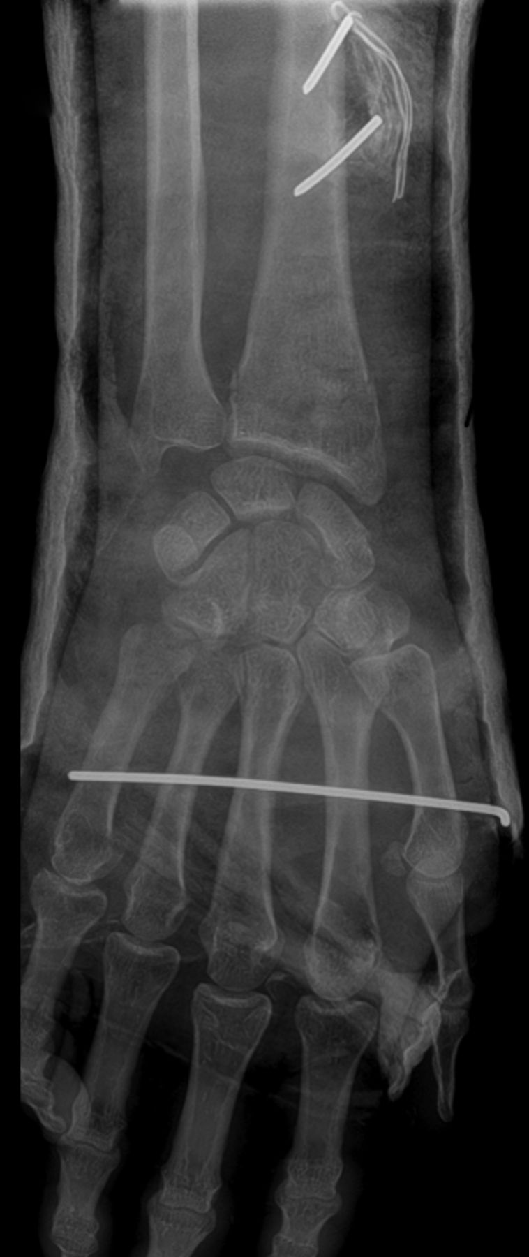

Figs. 1a - 1b
Radiographs showing a) an anteroposterior view with two pins inserted proximal to the fracture site, providing a buttress to maintain alignment. Another pin is inserted in the second and third metacarpal bone, with the pin bent at the radial side. The b) lateral view shows pins which engaged both cortices of the bone, and remained out of the skin
Fluoroscopy was used intra-operatively to confirm the reduction and placement of the pins. We took a control radiograph to assess possible loss of reduction within the first week of operation. The patients were followed up at one, three and six weeks, three and six months, and one and two years following hospital discharge. Radiological review was undertaken within the first week of surgery, at six weeks and one year, by measuring the radial inclination, radial tilt, and radial height. At the third month, sixth month, first year, and second year, we measured the range of movement (ROM) (flexion, extension, supination, and pronation) using a goniometer. Functional evaluation was carried out at the six-month and first-year follow-up visit using Solgaard’s modification9 of the scoring system described by Gartland and Werley.10 Evaluation of radiographic and functional results were completed by one senior physician (ARM).
Once the plaster was wet and had changed colour, it was considered to be infected. Thus, oral antibiotics were administered in order to avoid potential complications, including pin loosening, until the drainage issue had been resolved.
Pins were removed at the third week post-operatively, with initial cast removal followed by the application of a new short arm cast for an additional three weeks.
At six weeks, another radiograph was taken to assess the union, and decisions were made regarding the time of cast removal (delayed until the eighth week if necessary). Physical therapy was started after cast removal to return wrist movement and strength.
Results
We collected complete data from 54 patients with unstable fractures of the distal radius whom we successfully followed for one year. A total of 19 patients refused to participate in the study after removal of the pin and plaster. In total, 36 fractures were classified as type A and 18 were classified as type C fractures, according to the AO classification. All 54 patients were followed up for at least 12 months. The demographic results are summarised in Table I.
Post-operative radiographic results within the first week of surgery showed that the mean radial inclination was 23.7° (22° to 26°), the mean radial tilt was 10.6° (8° to 12°) of volar flexion, and the mean radial height was 10.2 mm (9 to 13). At six-week follow-up, visit the mean radial inclination was 24.5° (22° to 28°), radial tilt into volar flexion was 10.6° (8° to 12°), and the mean radial height was 10.2 mm (9 to 12). At one-year follow-up, the mean radial inclination, radial tilt, and radial height were 24.5° (22° to 28°), 10.1° (8° to 11°), and 10.2 mm (9 to 11), respectively (Table II).
Table II
Radiographic results. Data are presented as means with ranges
| Pre-operative n = 73 | Post-operative n = 73 | Six-wk follow-up (n = 73) | One-yr follow-up (n = 54) | |
|---|---|---|---|---|
| Mean radial inclination (°) | 16.7 (13 to 20) | 23.7 (22 to 26) | 24.48 (22 to 28) | 24.53 (22 to 28) |
| Mean radial tilt (°) | -24.9 (-21 to -29) | 10.6 (8 to 12) | 10.6 (8 to 12) | 10.1 (8 to 11) |
The ROM in the injured side compared with that in the opposite side at the three-month visit showed that the mean loss of flexion was 14° (10° to 22°), extension was 16.2° (9° to 21°), pronation was 7.7° (5° to 11°), and supination was 12.8° (4° to 16°). At the first-year follow-up visit the mean loss of flexion, extension, pronation, and supination were 6.7° (2° to 10°), 7.3° (2° to 12°), 0° (-3° to 2°) and 8° (3° to 11°), respectively (Table III).
Table III
Loss of range of movement (°) in the injured wrist compared with the uninjured wrist. Data are presented as mean differences between two wrists, with ranges
| Three mths n = 64 | Six mths n = 63 | One yr n = 54 | Two yrs n = 19 | |
|---|---|---|---|---|
| Flexion | 14 (10 to 22) | 7 (5 to 10) | 6.7 (2 to 10) | 6.5 (2 to 9) |
| Extension | 16.2 (9 to 21) | 8 (5 to 13) | 7.3 (2 to 12) | 7.2 (2 to 10) |
| Pronation | 7.7 (5 to 11) | 2.7 (-2 to 5) | 0 (-3 to 2) | 0.2 (-1 to 2) |
| Supination | 12.8 (4 to 16) | 8.5 (4 to 10) | 8 (3 to 11) | 7.8 (3 to 9) |
Functional evaluation (using Solgaard’s modification of the scoring system described by Gartland and Werley) estimated 26 patients with excellent results, good results in 25 patients, fair results in three patients, and no patient had a poor end result at the first-year follow-up visit (Table IV). All of the fractures healed in our series. Once physical therapy was completed, all patients achieved full range of active finger movement, with no pain and stiffness. There were three cases of pin tract drainage, which were treated successfully with empiric oral antibiotic, and earlier removal of the pin and plaster was not necessary. There were no cases of pin loosening. Three out of 54 patients suffered from radial paresthesis at the time of cast removal. All of them resolved at the six-month follow-up. No cases of median nerve compression or carpal tunnel syndrome were recorded in our series.
Table IV
Functional assessments using Solgaard’s modification of the scoring system described by Gartland and Werley
| Six mths follow-up | One yr follow-up | |
|---|---|---|
| Excellent | 29 | 26 |
| Good | 27 | 25 |
| Fair | 7 | 3 |
| Poor | 0 | 0 |
Discussion
There are several operative techniques available to achieve a congruent and stable articular surface and sufficient support for fractures of the distal radius.3,5,11-13 One such technique is percutaneous pinning with closed reduction followed by cast immobilisation. Percutaneous pinning could provide more stability in addition to sufficient support, compared with closed reduction and cast immobilisation alone.14 There are several studies reporting the outcomes of conventional manipulation and casting in comparison with other treatment options. Casting alone may not be enough to maintain anatomical position and reduction, leading to poorer functional and cosmetic results.15 However, in a study conducted by Azzopardi,16 it was shown that percutaneous pinning does not provide better clinical outcomes than cast immobilisation alone. The authors used two crossed Kirschner (K-) wires through the styloid process of the radius and through either Lister’s tubercle or the dorso-ulnar border of the distal fragment. However, in our study, we have used two pins in the fracture site and another one in the metacarpal bone, which were incorporated into the cast for more stability and support. This technique is both less invasive and technically demanding than open surgery. It is also suitable for reducing extra-articular fractures without intra-articular instability and metaphysical comminution, and in patients with good bone quality.3,6,8,11 Various pinning techniques have been described involving pinning through the radial styloid, crossed pins across the fracture site, or intrafocal pinning through the fracture site.3,12 Several complications have been mentioned; some are associated with the injury itself, and others result from treatment. Cooney et al17 categorised them into nine major types: compression neuropathy, arthrosis, malunion, tendon rupture, Volkmann’s ischemic contracture, arthrofibrosis of the fingers, shoulder–hand syndrome, unrecognised associated injuries, and complications of fixation.
We consider that our modified technique of pin in plaster can efficiently restore radial tilt and prevent radial shortening with good functional results, with fewer or comparable complications than those of other published techniques. The most common complications of percutaneous pinning are pin tract infection and radial nerve irritation.11,12 There were three cases of pin tract infection in our series, all of which were treated successfully with oral antibiotic therapy. There were no cases of pin loosening in our series, perhaps due to the application of antibiotics as mentioned above, when the plaster was wet. Three patients in our series suffered from radial nerve irritation. All three cases were treated early in the study. We took care to avoid injury to the surrounding soft tissues and superficial nerves using a 1 cm incision on the posterolateral aspect of the forearm before pin insertion, and protecting the nerve from irritation by cast moulding. Allain18 performed a trial study on 60 patients with dorsally displaced extra-articular or noncomminuted intra-articular fractures of the distal radius who received trans-styloid pin fixation with either one or six weeks of cast immobilisation, and reported one case of superficial infection, no deep infections, four cases of paraesthesia and/or hypoaesthesia in the radial nerve region, one case of tendon rupture of the extensor pollicis brevis and abductor pollicis longus, and one anterior pin migration. In another trial study performed by Azzopardi,16 one out of 27 patients suffered a pin tract infection with no tendon or neurovascular injuries reported. Lenoble19 compared trans-styloid and Kapandji pinning fixation, with eight cases (14.8%) of radial nerve irritation in the Kapandji group and three (7.1%) in the trans-styloid group. There were no tendon or vascular complications or median nerve dysfunction reported. Superficial infection was reported in three cases (5.5%) of the Kapandji group and in only one (2.3%) of the trans-styloid group. In another study of 140 children under the age of 16 who underwent percutaneous K-wire insertion either through the radial styloid or Lister’s tubercle, the rate of superficial pin tract infection was 5.7%, and medial and ulnar neuropraxia occurred in 1.4% of cases.20 Another prospective trial of Kapandji versus percutaneous extra-focal fixation revealed an 18% incidence of reflex sympathetic dystrophy in the Kapandji group and 8.6% in the extra-focal pinning group.21
Green22 described a type of pin-in-plaster technique in which the distal pin is inserted into the second and third metacarpals and the proximal pin is inserted through the ulna at least 6 cm from the tip of the olecranon, both covered by the sterile short arm cast. We applied a short arm cast to achieve immobilisation and more stability, therefore patients began hand movement from the first day after operation and were able to perform their routines with no finger or elbow stiffness. In addition, follow-up examinations such as radiographs are easier, relatively speaking, in a short arm cast compared with a long arm cast.
The strength of the present study is the use of the same technique in patients from young to old, with potentially different bone quality. However, it could be considered a limitation that we had no patients over 73 years of age. One explanation for this is that older patients may be reluctant to undergo surgery and prefer to follow their own traditional treatment, such as fixation of fracture or application of a poultice in order to enable relief from pain and symptoms. A further limitation of our study is a loss of follow-up over time, which may jeopardise the validity of both the radiological and functional outcome conclusions. This loss to follow-up could be attributed to the difficulty for patients of long-distance travel, as the majority of patients who left the study were from rural areas.
In conclusion, our study supports pin-in-plaster fixation as an excellent technique for the treatment of unstable and comminuted fractures of the distal radius. The technique is less invasive and technically easier than other more complex interventions, and efficiently restores anatomic congruity and maintains reduction.
1 Nellans KW , KowalskiE, ChungKC. The epidemiology of distal radius fractures. Hand Clin2012;28:113–125.CrossrefPubMed Google Scholar
2 Mellstrand C , PetterssonHJ, TornqvistH, PonzerS. The operative treatment of fractures of the distal radius is increasing: results from a nationwide Swedish study. Bone Joint J2014;96-B;963–969.CrossrefPubMed Google Scholar
3 Shin EK , JupiterJB. Current concepts in the management of distal radius fractures. Acta Chir Orthop Traumatol Cech2007;74:233–246.PubMed Google Scholar
4 Mirghasemi AR , LeeDJ, RahimiN, RashidiniaS, ElfarJC. Distal Radioulnar Joint Instability. Geriatr Orthop Surg Rehabil2015;6:225–229.CrossrefPubMed Google Scholar
5 Liporace FA , AdamsMR, CapoJT, KovalKJ. Distal radius fractures. J Orthop Trauma2009;23:739–748.CrossrefPubMed Google Scholar
6 Handoll HH , VaghelaMV, MadhokR. Percutaneous pinning for treating distal radial fractures in adults. Cochrane Database Syst Rev2007;3:CD006080.CrossrefPubMed Google Scholar
7 Kivi MM , AsadiK, MotlaghKH, ShakibaM. Distal radius fracture, a comparison between Closed reduction and Long arm cast vs. Closed reduction and Percutaneous pinning and Short arm cast. Shiraz E Medical Journal2011;12:155–161. Google Scholar
8 Chen CE , JuhnRJ, KoJY. Treatment of distal radius fractures with percutaneous pinning and pin-in-plaster. Hand (N Y)2008;3:245–250.CrossrefPubMed Google Scholar
9 Solgaard S . Function after distal radius fracture. Acta Orthop Scand1988;59:39–42.CrossrefPubMed Google Scholar
10 Gartland JJ , WerleyCW. Evaluation of healed Colles' fractures. J Bone Joint Surg [Am]1951;33-A:895–907.PubMed Google Scholar
11 Vasenius J . Operative treatment of distal radius fractures. Scand J Surg2008;97:290–296.CrossrefPubMed Google Scholar
12 Henry MH . Distal radius fractures: current concepts. J Hand Surg Am2008;33:1215–1227.CrossrefPubMed Google Scholar
13 Inglis M , McClellandB, SutherlandLM, CundyPJ. Synthetic versus plaster of Paris casts in the treatment of fractures of the forearm in children: a randomised trial of clinical outcomes and patient satisfaction. Bone Joint J2013;95-B:1285–1289.CrossrefPubMed Google Scholar
14 Das AK , SundaramN, PrasadTG, ThanhaveluSK. Percutaneous pinning for non-comminuted extra-articular fractures of distal radius. Indian J Orthop2011;45:422–426.CrossrefPubMed Google Scholar
15 Howard PW , StewartHD, HindRE, BurkeFD. External fixation or plaster for severely displaced comminuted Colles’ fractures? A prospective study of anatomical and functional results. J Bone Joint Surg [Br]1989;71-B:68–73. Google Scholar
16 Azzopardi T , EhrendorferS, CoultonT, AbelaM. Unstable extra-articular fractures of the distal radius: a prospective, randomised study of immobilisation in a cast versus supplementary percutaneous pinning. J Bone Joint Surg [Br]2005;87-B:837–840.CrossrefPubMed Google Scholar
17 Cooney WP III , DobynsJH, LinscheidRL. Complications of Colles’ fractures. J Bone Joint Surg [Am]1980;62-A:613–619. Google Scholar
18 Allain J , le GuillouxP, Le MouëlS, GoutallierD. Trans-styloid fixation of fractures of the distal radius. A prospective randomized comparison between 6- and 1-week postoperative immobilization in 60 fractures. Acta Orthop Scand1999;70:119–123.CrossrefPubMed Google Scholar
19 Lenoble E , DumontierC, GoutallierD, ApoilA. Fracture of the distal radius. A prospective comparison between trans-styloid and Kapandji fixations. J Bone Joint Surg [Br]1995;77-B:562–567.PubMed Google Scholar
20 Choi KY , ChanWS, LamTP, ChengJC. Percutaneous Kirschner-wire pinning for severely displaced distal radial fractures in children. A report of 157 cases. J Bone Joint Surg [Br]1995;77-B:797–801.PubMed Google Scholar
21 Mirhamidi SM , BayatFM. A prospective comparison between Kapandji and percutaneous extra-focal fixation in extra articular distal radius fractures. Int J Clin Exp Med2013;6:133–139.PubMed Google Scholar
22 Green DP . Pins and plaster treatment of comminuted fractures of the distal end of the radius. J Bone Joint Surg [Am]1975;57-A:304–310.PubMed Google Scholar
Funding statement:
None declared
Author contributions:
S. A. Mirghasemi: Data collection, performed surgeries, research team manager.
S. Rashidinia: Writing the paper, data analysis and collection.
M. S. Sadeghi: Writing the paper, data analysis and collection.
M. Talebizadeh: Searching the articles, data analysis.
N. Rahimi: Data collection.
ICMJE Conflict of Interest:
None declared
©2015 Mirghasemi et al. This is an open-access article distributed under the terms of the Creative Commons Attributions licence (CC-BY-NC), which permits unrestricted use, distribution, and reproduction in any medium, but not for commercial gain, provided the original author and source are credited.
